Αρχείο:Yellow mite (Tydeidae) Lorryia formosa 2 edit.jpg

Πρωτότυπο αρχείο (2.100 × 2.560 εικονοστοιχεία, μέγεθος αρχείου: 1,63 MB, τύπος MIME: image/jpeg)
Σύνοψη
| Περιγραφή |
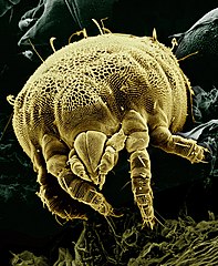
English: Historically, mites have been difficult to study because of their minute size. But now, ARS scientists are freezing mites in their tracks and using scanning electron microscopy to observe them in detail. Here a yellow mite, Lorryia formosa, commonly found on citrus plants, is shown among some fungi. False color. Magnified about 850x. |
|||
| Ημερομηνία | ||||
| Πηγή |
|
|||
| Δημιουργός |
Photo by Eric Erbe; digital colorization by Chris Pooley. Edited by Fir0002 |
|||
| Άδεια (Επαναχρησιμοποίηση αυτού του αρχείου) |
From Christopher Pooley to brian0918, March 22, 2005 3:19 PM: "Thank you for your interest in our images. All of the micrographs on the web site are in the public domain and can be freely used. Proper accreditation would be "Erbe, Pooley: USDA, ARS, EMU". High Resolution copies of the web images are available on our FTP site ftp://198.77.171.17/pub/"
|
|||
| άλλες εκδόσεις |
|
,_Lorryia_formosa_2.jpg)
_Lorryia_formosa_2_edit.jpg)
|
_Lorryia_formosa_2_edit.jpg)
_Lorryia_formosa_2_edit.jpg)
_Lorryia_formosa_2_edit.jpg)
Ιστορικό αρχείου
Πατήστε σε μια ημερομηνία/ώρα για να δείτε το αρχείο όπως εμφανιζόταν εκείνη την χρονική στιγμή.
| Ημερομηνία/Ώρα | Μικρογραφία | Διαστάσεις | Χρήστης | Σχόλιο | |
|---|---|---|---|---|---|
| τρέχον | 06:56, 18 Ιουλίου 2006 | | 2.100 × 2.560 (1,63 MB) | Fir0002 | Historically, mites have been difficult to study because of their minute size. But now, ARS scientists are freezing mites in their tracks and using scanning electron microscopy to observe them in detail. Here a yellow mite, Lorryia formosa, commonly found |
| 06:45, 18 Ιουλίου 2006 |  | 2.100 × 3.000 (1,85 MB) | Fir0002 | Historically, mites have been difficult to study because of their minute size. But now, ARS scientists are freezing mites in their tracks and using scanning electron microscopy to observe them in detail. Here a yellow mite, Lorryia formosa, commonly found |
Χρήση αρχείου
Η ακόλουθη σελίδα χρησιμοποιεί προς αυτό το αρχείο:
Καθολική χρήση αρχείου
Τα ακόλουθα άλλα wiki χρησιμοποιούν αυτό το αρχείο:
- Χρήση σε ar.wikipedia.org
- Χρήση σε ast.wikipedia.org
- Χρήση σε bn.wikipedia.org
- Χρήση σε da.wikipedia.org
- Χρήση σε el.wikipedia.org
- Χρήση σε en.wikipedia.org
- Nature
- Chelicerata
- Mite
- Wikipedia:Featured pictures thumbs/05
- Wikipedia:Picture of the day/September 2006
- Wikipedia:Featured picture candidates/July-2006
- Wikipedia:Featured picture candidates/Yellow mite
- Wikipedia:Wikipedia Signpost/2006-07-24/Features and admins
- Wikipedia:WikiProject Spiders/Articles
- User:Fir0002/Fir0002 gallery/Featured Pictures edits
- Wikipedia:Picture of the day/September 20, 2006
- Wikipedia:POTD/September 20, 2006
- Wikipedia:POTD column/September 20, 2006
- Wikipedia:POTD row/September 20, 2006
- User talk:Brian0918/Archive 24
- Wikipedia:WikiProject Arthropods/POTD
- Microfauna
- Wikipedia:Featured pictures/Animals/Arachnids
- User:Xophist/s3
- User:Siva.tecz/sandbox
- User:Armanaziz/Nature
- Wikipedia:Wikipedia Signpost/Single/2006-07-24
- User:Kait.Snow/Microfauna
- Χρήση σε en.wikibooks.org
- Χρήση σε es.wikipedia.org
- Χρήση σε fa.wikipedia.org
- طبیعت
- ویکیپدیا:نگارههای برگزیده/حشرات
- ویکیپدیا:نگاره روز/دسامبر ۲۰۱۳
- ویکیپدیا:گزیدن نگاره برگزیده/ژوئن-۲۰۱۳
- هیره زرد
- ویکیپدیا:گزیدن نگاره برگزیده/Yellow mite (Tydeidae) Lorryia formosa 2 edit.jpg
- الگو:نر/2013-12-17
- الگو:نر محافظت شده/2013-12-17
- بحث کاربر:Alborzagros/بایگانی ۱۵
- هیره
- ریزجانوران
- ریززیاگان
- Χρήση σε fr.wikipedia.org
- Χρήση σε gv.wikipedia.org
_Lorryia_formosa_2_edit.jpg)
Δείτε περισσότερη καθολική χρήση αυτού του αρχείου.
_Lorryia_formosa_2_edit.jpg)
_Lorryia_formosa_2_edit.jpg)